Biometrics Blog
Permanent link for Research Review: "Enhancing Imaging Anatomy Competency: Integrating Digital Imaging and Communications in Medicine (DICOM) Viewers Into the Anatomy Lab Experience" on January 24, 2025
Biometrics research continues to play a critical role in how we make advancements in education. In a recent study, researchers tested whether or not viewing Digital Imaging Communications in Medicine (DICOM) helped medical students do better, and feel more confident on tests.
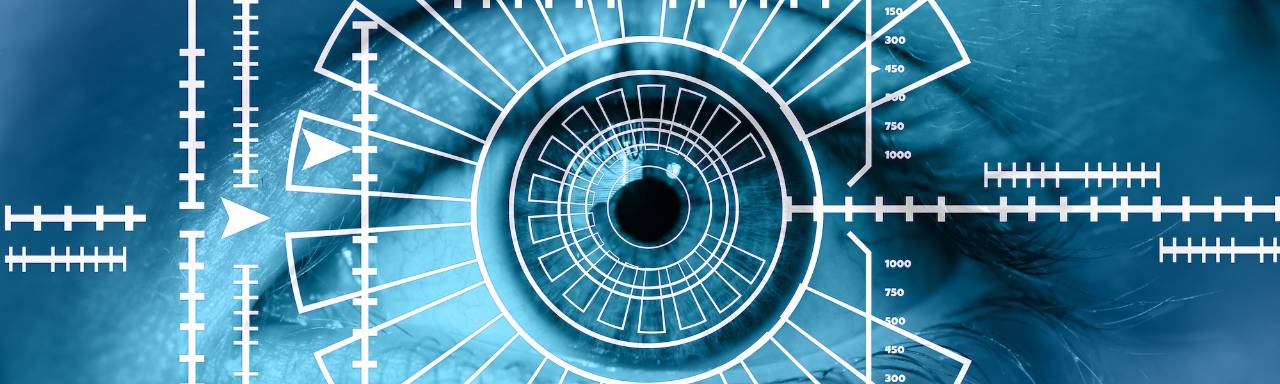
Many medical students are only given the opportunity to view static anatomy prior to testing in medical school, which leads both educators and researchers wondering if students are able to systematically interpret scan images, or are simply memorizing the patterns shown to them. By using eye-tracking software, researchers can review conscious and subconscious behavior of students, analyzing their gaze patterns and visual-spatial evaluations (Worley Et al., 2).
How was this study conducted:
- Nine first year medical students (the test group) were exposed to DICOM software in their anatomy course, and ten second year medical students (the control group) were exposed to static images in a powerpoint presentation in their anatomy course.
- The students were given a pre-quiz survey asking them to rank their “confidence in identifying anatomical structures in cardiothoracic imaging using a 10-point Likert scale (1 = not confident at all; 10 = very confident)” (Worley Et al., 2).
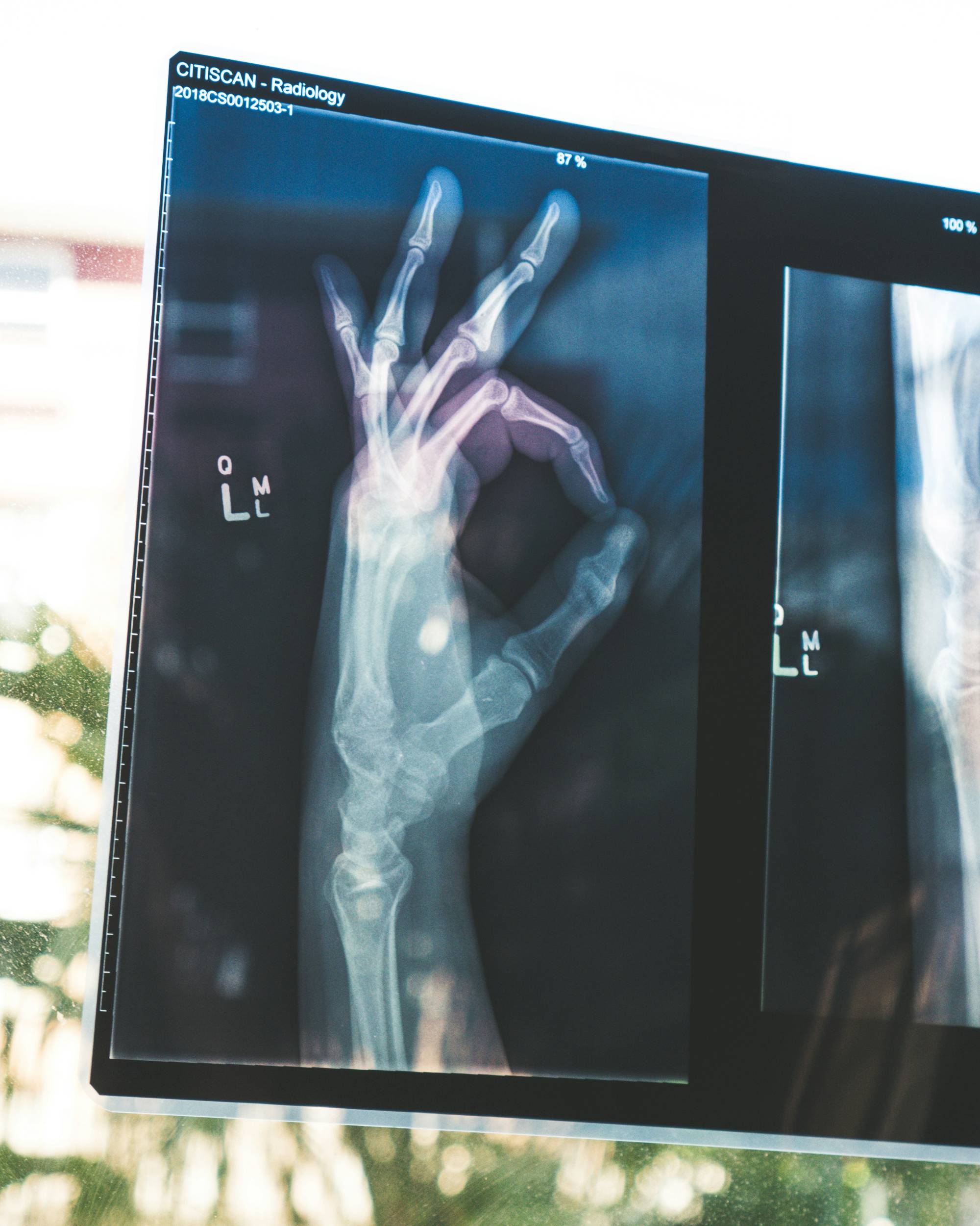
- They were then asked to identify anatomic structures on 15 digital radiographic images, while tracking their eye-movement, focusing on saccade peak velocity and fixation duration.
Findings:
The study found that the first year students exposed to DICOM images did significantly better on the test than the second year students who were not. Despite this, there were no significantly different reported levels of confidence, saccade peak velocities, or fixation durations. As a result, no strong conclusions can be made about whether or not the test group was significantly more capable of reviewing the images or not, but this may also be due to the small sample size of the test group.
While the curriculum change from Power Point images to DICOM software hasn’t proven to create extremely remarkable changes in medical students’ education, it does show some level of improvement in identifying anatomical structures. Despite this, a major limitation of the study is that the second year medical students were provided their Power Point lesson nine months prior to conducting this study, whereas the first year students utilized the DICOM software only two to five months prior to conducting the study. But, this may be rendered moot due to the second year students’ longer exposure to experience with anatomy due to their sophomore status in the program.
Lessons from this Publication
- It is important to have a large sample size when doing biometric research, as it allows for larger generalizations to be made- which is particularly important in educational contexts for educators to apply the findings of new research to their curriculums and classrooms.
- The researchers also asked participants how long they spent studying for the quiz, but due to the open-ended nature of their question, they weren’t able to derive any meaningful results. This indicates a need for objective survey questions that can produce distinct results when aiming to create quantifiable data points.
- It is important to consider extraneous variables in research. In this publication, the researchers assume that the first years’ more recent exposure to anatomy class “cancels out” the second years’ longer exposure to anatomy, but it is possible that these variables don’t have equal effects on the test subjects.
What does this mean for us?
This study emphasizes the importance of educational uses for biometrics. While statistically insignificant in this study, this method has the opportunity to provide valuable insights when students are required to analyze imagery by watching their patterns, areas of interest, and time spent in each area of interest.
Click here to read the full article.
Worley L, Colley M A, Rodriguez C C, et al. (September 07, 2024) Enhancing Imaging Anatomy Competency: Integrating Digital Imaging and Communications in Medicine (DICOM) Viewers Into the Anatomy Lab Experience. Cureus 16(9): e68878. doi:10.7759/cureus.68878
Categories:
Biometrics Research
Posted
by
Georgia Hessel
on
Permanent link for Research Review: "Enhancing Imaging Anatomy Competency: Integrating Digital Imaging and Communications in Medicine (DICOM) Viewers Into the Anatomy Lab Experience" on January 24, 2025.