Plastination in BMS
[1536586053].jpg)
Human placenta & umbilical cord
GVSU Plastination Lab News-2018
For readers who have not been exposed to the term, plastination refers to the process of impregnating animal or plant tissues with a variety of plastic or silicone products to render the tissues odor-free and permanently preserved. The process was developed in Germany by Dr. Gunther von Hagens, and made popular through his many Body Worlds exhibits around the world.
History
In fall, 2007 biologist Bruce Ostrow spent a sabbatical semester at the University of Michigan plastination lab, where he learned several plastination techniques and produced some plastinates for use in BIO and BMS courses. His efforts to establish a plastination lab at GVSU were unsuccessful until CLAS Dean Frederick Antzack provided funds for this venture in the spring of 2013. During July 2013 anatomist Tim Strickler completed a workshop at the University of Toledo to learn a variety of plastination techniques, and he and Bruce began to assemble the materials required to begin plastinating at GVSU. 2013 saw the demise of the plastination lab at the University of Michigan, with the result that our new lab became the only university plastination facility in the state. Plastination of pig hearts and kidneys began in 217 Padnos Hall in September 2013, and to date we have produced hundreds of plastinates for use in GVSU courses. In June 2014, anatomist Dawn Richiert and anatomy lab supervisor Kim Wieber also completed the University of Toledo workshop. The lab moved to a new location in 263C Padnos Hall in the fall of 2016.
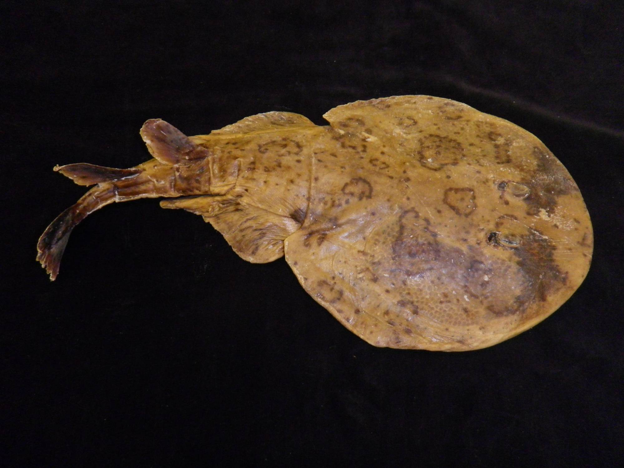
Lesser Electric Ray – Narcine bancroftii - one of our plastinated fish
The Process
The plastination process begins with dissection of the specimen, usually carried out by students (undergraduate and graduate) who have proven talents in dissection, and who find the preparation and processing of plastinates a fascinating activity. The specimen is then washed and bleached, and dehydrated in acetone. The dehydrated specimen is submerged in a vacuum chamber filled with liquid silicone and acetone is drawn out of the specimen under vacuum. As the acetone is removed, silicone is drawn into the tissues until all of the acetone has been replaced by silicone. The specimens are then removed from the silicone tank and the excess silicone is allowed to drain over a period of days, weeks, or months. Finally, the specimens are posed in what will be their final positions, and treated with a catalyst, which causes the silicone to set up and become dry to the touch. They are then ready to be used by students in our anatomy and biology labs and in several art courses. Carefully handled, these specimens will last for decades, and their use is completely without the mess and exposure to dangerous chemicals that are usually associated with wet specimens.
There are two silicone plastination techniques- one done at -20°C or colder, and the other at room temperature. We are using the room temperature technique, which produces specimens equal in quality to the cold technique and also uses much less energy. In addition, the silicone polymer remains fluid and can be used repeatedly. The cold mixture will set up at room temperature, and therefore has to be kept cold, whether or not it is being used to process tissue.
Plastinates produced
Plastinated specimens produced in the GVSU lab in the past five years range from a variety of vertebrates used in several biology courses (fish, salamanders, frogs, turtles, cats and fetal pigs) to human specimens acquired from the University of Michigan for this purpose. MSU does not permit us to retain human specimens beyond their normal two-year period of use, so the human material from the cadavers we receive from them cannot be plastinated. We have also managed to plastinate a few invertebrates and a few plant specimens. With the addition of three large vacuum tanks in 2014 and 2015, we are now able to plastinate rather large items, including a full-term 2-headed calf. We have completed the plastination of the musculoskeletal portions of two of our seven University of Michigan cadavers in addition to a variety of organ and joint specimens. With the cooperation of the MSU College of Veterinary Medicine, we have provided dog and pony plastinates to both MSU and the St Georges University School of Veterinary Medicine in Grenada, and have plastinated pathological specimens of intestine for a study of swine ileitis.
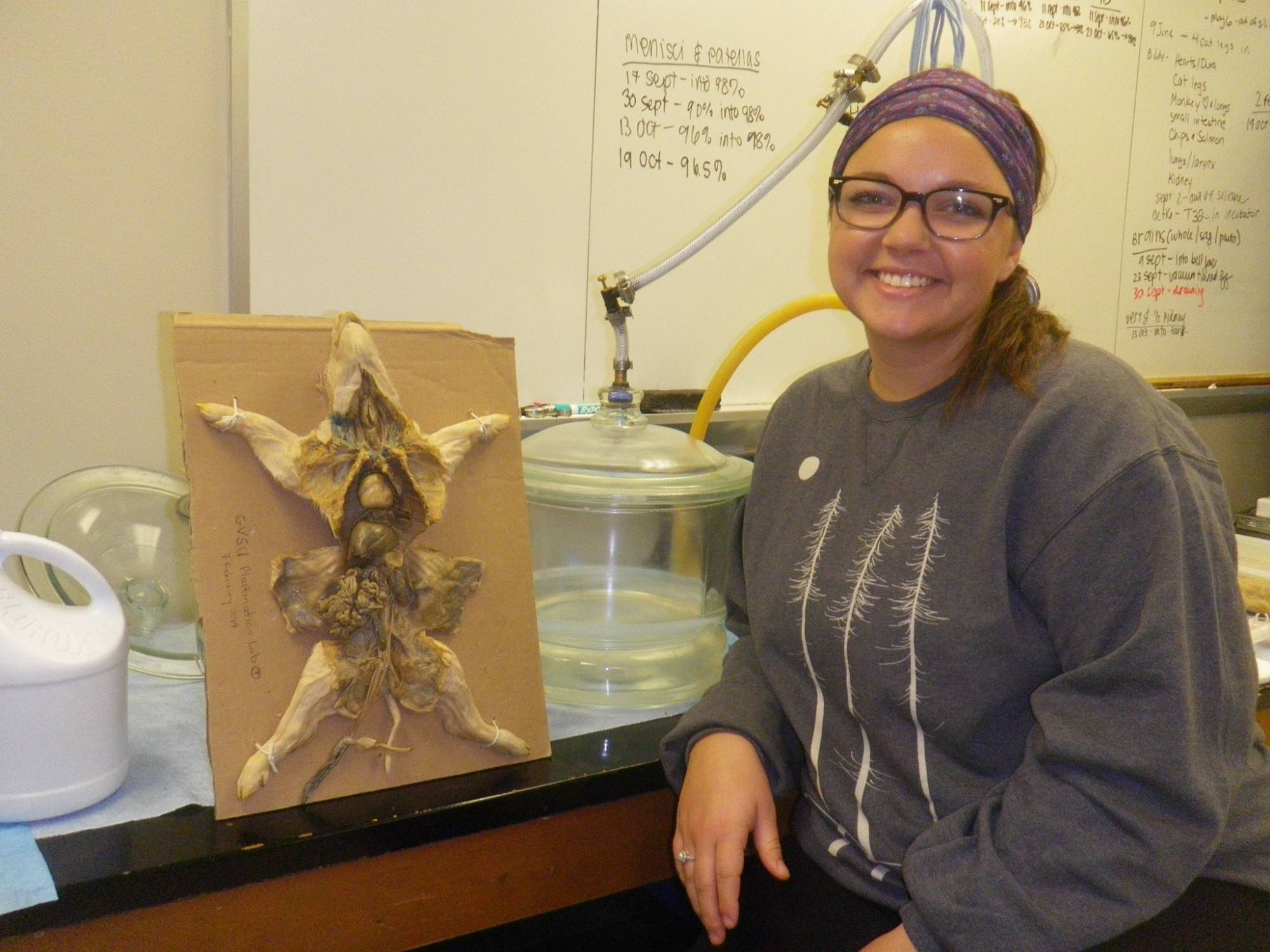
Lauren Begg, MHS Graduate Student dissector & fetal pig used in BIO 121
Recycling and energy efficiency
Because the plastination process uses large amounts of acetone and alcohol, one of the most important pieces of equipment in the lab is our recycler, which is able to reclaim acetone, ethanol and propanol. As the specimens are dehydrated in acetone, the water content of the solution increases. We start the dehydration process with 98% acetone (the concentration of the end product from the recycler), and when it declines to 80-85% we remove it for recycling and refill the dehydration container with fresh 98% acetone. Since the start of the lab in September 2013, we have reclaimed over 1500 gallons of acetone and 50 gallons of ethanol. We have also made our recycler available to the chemistry department for their recycling needs, primarily acetone and isopropanol.
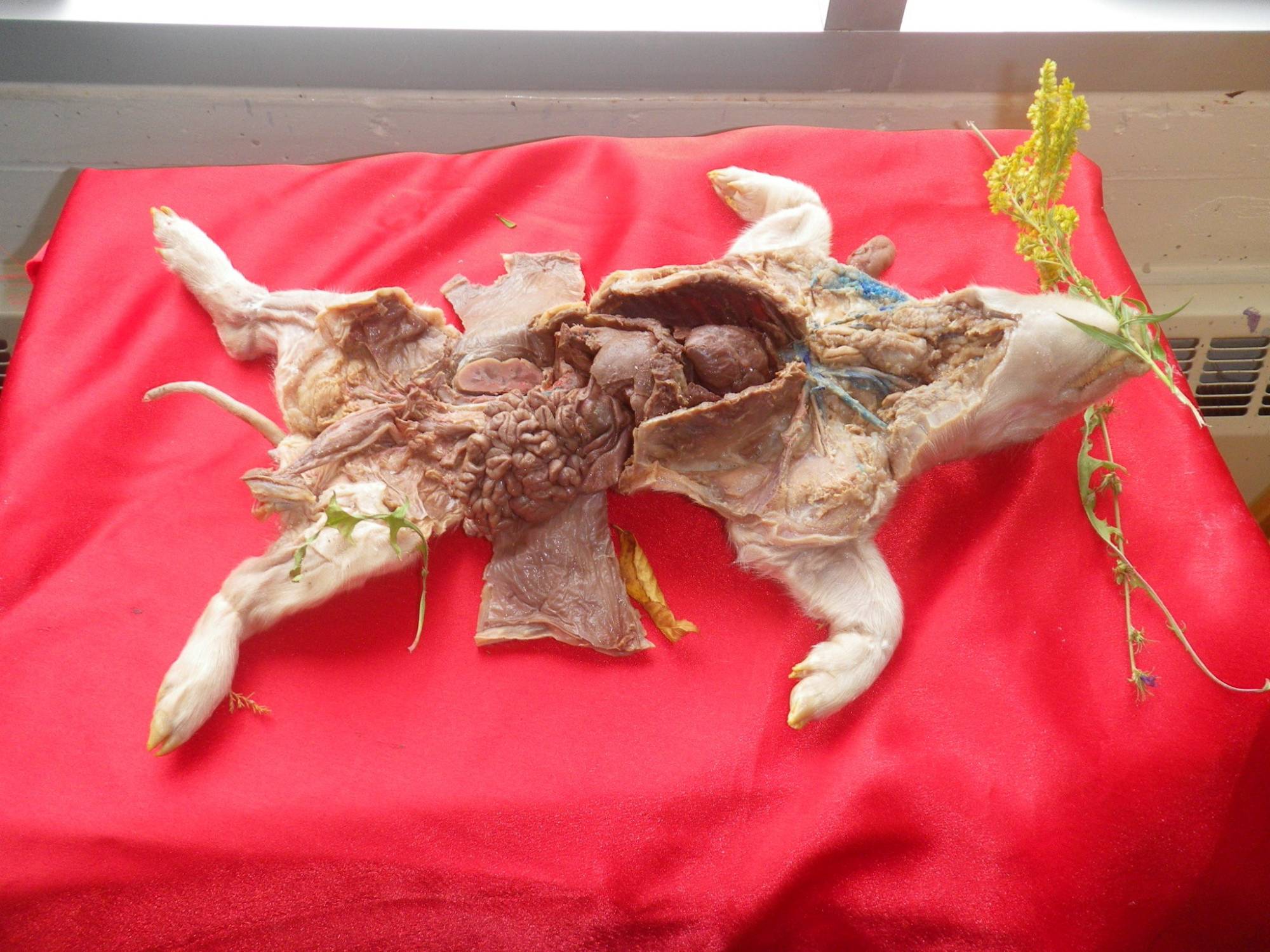
Lauren’s fetal pig in the Art Studio- some of our non-human plastinates are used as models for students in several ART courses